
Ever stared at a ELISA kit and felt like you were deciphering a secret code?
We’ve all been there—pipette in hand, plate glistening, and that nagging question of “Did I set this up right?” especially when your experiment could mean a breakthrough for a research lab or a diagnostic test.
That uneasy feeling is why we’re breaking down the elisa procedure steps in a way that feels less like a textbook and more like a chat over coffee.
First, we’ll walk through the sample preparation, because getting a clean, well‑diluted sample is the foundation of any reliable assay. Imagine you’re mixing a smoothie; too much fruit or too little liquid throws off the texture, same with your samples.
Next comes the plate coating. Whether you’re using a pre‑coated plate or applying your own capture antibody, the key is a gentle, even spread—think of it as painting a wall, you want smooth coverage without drips.
Then we add the blocking step. This is the unsung hero that stops background noise, kind of like putting a rug on a squeaky floor.
After blocking, the samples and standards are added. Here, timing matters; you’ll want to let each incubation sit just long enough to bind without over‑exposing.
Washing follows, and it’s where many labs slip—skip it and you’ll get a mess of false positives. A good rule is to tap the plate gently, add wash buffer, and repeat three times.
The detection antibody comes next, delivering that fluorescent or colorimetric signal we all chase. Think of it as the spotlight on your stage.
Finally, the substrate reaction and reading on a microplate reader seal the deal. A quick glance at the absorbance values tells you whether your hypothesis holds.
Throughout, we’ll sprinkle practical tips—like using a calibrated micropipette (check our guide on how to use a micropipette) and keeping everything at the right temperature.
By the end, you’ll have a clear roadmap that turns the elisa procedure steps from a daunting checklist into a smooth, repeatable workflow.
Ready to demystify ELISA and get confident results? Let’s dive in.
TL;DR
The ELISA procedure steps, from sample prep to reading absorbance, are broken down into clear, coffee‑chat style guidance so you can avoid common pitfalls and get reliable results every time.
Follow our practical tips, like calibrated pipetting and proper blocking, to turn a daunting checklist into a smooth, repeatable workflow.
Step 1: Sample Collection and Preparation
Before the ELISA plate ever sees a drop of sample, you’ve got to treat the material like a fragile heirloom. A sloppy collection will ruin everything downstream, no matter how perfect your washing steps are.
First thing: decide what matrix you’re working with—serum, plasma, cell culture supernatant, or tissue lysate. Each one has its own quirks. Serum, for example, tends to clot if you don’t add clot‑activator tubes, while tissue lysates need a protease inhibitor cocktail to keep proteins intact.
Collect the sample in a low‑bind microcentrifuge tube. Keep it on ice if you’re dealing with enzymes that degrade quickly. A quick spin at 10,000 × g for 5 minutes clears debris, leaving a clear supernatant that’s ready for dilution.
Now comes the dilution step. Think of it like sweetening a coffee—you want just enough water so the flavor isn’t too strong, but not so much that it’s watery. For most ELISA kits, a 1:2 to 1:10 dilution works, but always double‑check the kit’s recommended range.
Use a calibrated micropipette for every transfer. If you’re unsure how to get consistent volumes, our guide on how to use a micropipette walks you through setting the tip, zeroing, and avoiding air bubbles.
Don’t forget to label each tube clearly. A simple mistake—mixing up a control with a test sample—can cause you to waste an entire plate. Write the sample ID, dilution factor, and collection time on a waterproof label.
Once everything is labeled, store the aliquots at the temperature the kit recommends. Most proteins are stable at –20 °C for weeks, but cytokines often need –80 °C to avoid degradation.
Tip: if you’re running a high‑throughput screen, consider using a multichannel pipette or an automated liquid‑handling system. It speeds up the process and reduces pipetting error, which is a common source of variability in the elisa procedure steps.
Before you pour anything onto the plate, run a quick quality‑control check. Measure protein concentration with a spectrophotometer or use a BCA assay to confirm you’re in the kit’s dynamic range. If the reading is off, dilute or concentrate accordingly—there’s no point proceeding with a sample that’s out of range.
While you’re waiting for the samples to thaw or equilibrate, take a moment to think about the bigger picture. If your lab is a small startup, you’re probably wearing many hats—scientist, manager, and HR officer all at once.
That’s why it can be useful to have a quick reference on employee benefits. A practical guide on health insurance for small businesses can save you hours of Googling later.
And when you’ve nailed the wet‑lab side, don’t forget the dry‑lab side: making sure your research group shows up in search results. A platform like Rebelgrowth can help you build a solid online presence so collaborators and funders find you easily.

By treating the collection step with the same care you’d give a delicate experiment, you set the stage for reproducible results. Remember: clean, well‑labeled, properly stored samples are the foundation of any successful ELISA assay. That attention to detail pays off when the data finally come in.
Step 2: Coating the Plate with Capture Antibody
Alright, you’ve got your samples ready – now it’s time to give the plate something to hold onto. That’s where the capture antibody comes in, acting like a friendly net that grabs the target protein and keeps it from drifting away.
Does it feel a bit like painting a wall? Exactly. You want an even, thin layer that sticks without dripping. The trick is choosing the right buffer, concentration, and incubation conditions so the antibody sits nicely on the plastic.
Pick the right coating buffer
Most labs use a carbonate‑bicarbonate buffer (0.2 M, pH 9.4) because the high pH keeps the antibody soluble and promotes hydrophobic attachment to the polystyrene surface. If you’re working with a particularly delicate antibody, a phosphate‑buffered saline (pH 7.4) can be gentler, but you may need a slightly higher concentration to get the same binding efficiency.
Thermo Fisher’s guide on ELISA plate coating walks through the pros and cons of each buffer type, so it’s worth a quick glance if you’re unsure which one fits your assay Thermo Fisher’s guide on ELISA plate coating.
Once you’ve settled on a buffer, dissolve your capture antibody to a final concentration of 2–10 µg/mL. Too much antibody can cause “hooking” – the dreaded background signal that masks real results. A quick tip: start low (2 µg/mL) and run a small test plate to see how the signal‑to‑noise ratio looks.
Coating steps at a glance
1. Add 100 µL of the diluted capture antibody to each well.
2. Cover the plate with a sealing film to prevent evaporation.
3. Incubate at 4 °C overnight or at 37 °C for 1–2 hours if you’re short on time.
4. Gently discard the coating solution and wash three times with PBS‑Tween (0.05 %).
5. Immediately add blocking buffer (e.g., 1 % BSA) to keep the remaining surface from binding nonspecific proteins later on.
Sounds simple, but a couple of pitfalls can ruin the whole experiment. Have you ever forgotten to wash after coating? The leftover antibody can create high background that looks like a false positive. And if you skip the blocking step, even the cleanest coating will pick up stray proteins during later incubations.
Here’s a quick visual of the workflow – the video below walks through each step while a technician demonstrates the technique on a 96‑well plate.
After the video, take a moment to compare the three most common coating approaches. The table helps you decide which one matches your lab’s resources and the nature of your target antigen.
| Coating Method | Best For | Key Considerations |
|---|---|---|
| Passive adsorption (carbonate buffer) | Most antibodies, standard ELISA kits | Simple, low cost; watch out for orientation issues |
| Protein A/G‑pre‑coated plates | Antibodies that bind Protein A/G well (IgG subclasses) | Improves orientation; avoid for sandwich assays where cross‑reactivity could occur |
| Biotin‑streptavidin system | Biotinylated capture antibodies or small peptide antigens | Very strong, specific binding; requires extra biotinylation step |
When you pick a method, think about the downstream detection. If you plan to use a secondary antibody from a different species, a Protein A/G plate might unintentionally bind that secondary and raise background. In that case, stick with passive adsorption and fine‑tune the buffer pH.
One more thing: temperature matters. Overnight incubation at 4 °C often gives the most stable coating, but if you’re in a rush, a 1‑hour 37 °C step works fine – just be sure to verify the signal with a control well.
Finally, always run a “no‑antibody” control plate. It tells you how much background the plate itself contributes. If that number is high, you might need a different plate type or a more stringent blocking buffer. Thermo Fisher’s article on factors that affect ELISA signal explains why those controls are critical factors that affect ELISA signal.
Bottom line: a well‑coated plate is the foundation of a reliable ELISA. Take a few minutes to get the buffer, concentration, and incubation right, and you’ll save hours troubleshooting later. Ready to move on to the blocking step? Let’s keep the momentum going.
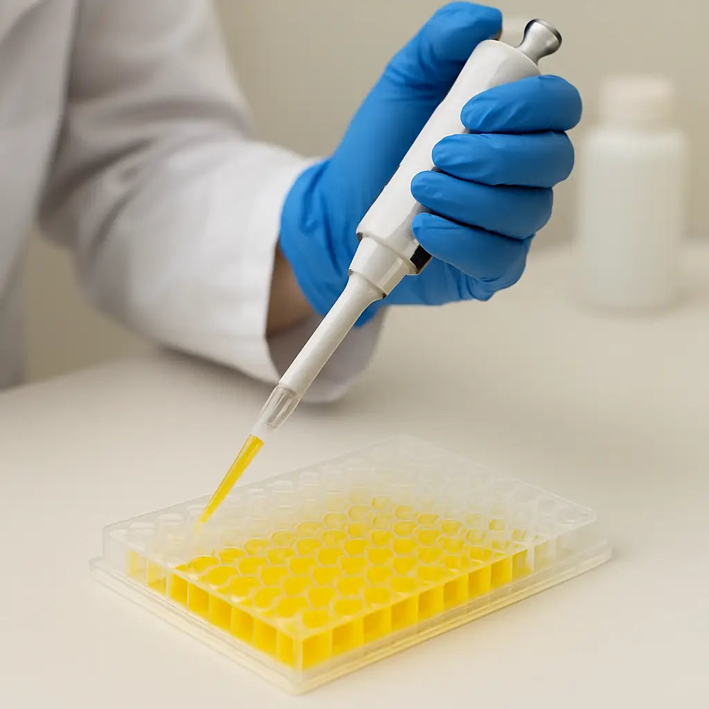
Step 3: Adding Samples and Standards
Now that your plate is coated and blocked, it’s time to actually put the biology in the wells. This is the step where the assay starts to feel like a real experiment instead of a set of instructions.
First thing’s first: make sure you have a fresh batch of standards prepared. Standards are just diluted versions of a known concentration of your target protein, and they give you the curve you’ll use to translate absorbance into actual amounts.
Preparing your standards
Grab the stock solution that came with the kit. If you’re using a custom antigen, you’ll need to know its exact concentration – a quick check with a spectrophotometer (if you have one) can save you a lot of guesswork later.
Typical ELISA kits call for a 7‑point, two‑fold dilution series. Start with the highest concentration (say 1000 pg/mL) and add an equal volume of diluent to make 500 pg/mL, then repeat until you reach the lowest point. Use low‑binding tubes so the protein doesn’t stick to the walls.
Tip: label each tube as you go. A simple “S1, S2…” on a sticky note prevents the classic “I think this is the 250 pg/mL but maybe it’s 125 pg/mL” moment.
Loading samples
Now turn to your prepared samples from Step 1. Remember the dilution you calculated earlier? If your sample is expected to sit somewhere in the middle of the standard curve, a 1:10 or 1:20 dilution is usually safe. For very low‑abundance targets, you might need to concentrate the sample or run it undiluted.
Using a calibrated pipette (yes, the one we talked about in the first step), add 100 µL of each standard or sample to the appropriate wells. It helps to work from the highest concentration down – that way you’re less likely to accidentally contaminate a low‑concentration well with a splash from a higher one.
Don’t forget the “blank” well that contains only diluent. This will be your zero point for background subtraction.
Tips to avoid common pitfalls
Ever wondered why some plates look perfect at the end and others are a mess of bubbles? One quick fix is to tap the plate gently on the bench after you add each 100 µL. The liquid will spread evenly and any trapped air will rise to the top.
If you’re working in a busy lab, set a timer for each incubation step. It’s easy to lose track and leave the samples on the plate for too long – that can lead to “hook effect” where the signal actually drops at high concentrations.
Another thing to watch: edge effects. The outer wells of a 96‑well plate can dry out faster, especially if you’re running the assay on a warm bench. If you have a lot of samples, consider filling the outer ring with buffer to keep the temperature uniform.
When you’re done loading, seal the plate with a plate sealer or a simple adhesive film. This prevents evaporation during the incubation and keeps the wells from pulling in dust.
Finally, give yourself a quick sanity check: glance at the plate, make sure each row has a logical order (standards decreasing, samples grouped together) and that no well is missing liquid. A brief visual scan now saves hours of troubleshooting later.
Once the plate is sealed, you’re ready for the incubation step that follows – the detection antibody will soon light up the spots where your target bound. Stay tuned, and you’ll see how a clean, well‑organized loading step makes the whole ELISA procedure steps feel like a smooth, repeatable workflow.
Step 4: Detection Antibody and Enzyme Conjugate
Okay, the plate is coated, blocked, and loaded – now the magic really begins. The detection antibody is the little spotlight that tells you where your target protein actually sits.
First thing you’ll notice is that the detection antibody usually comes pre‑conjugated to an enzyme, most often horseradish peroxidase (HRP) or alkaline phosphatase (AP). Those enzymes are the workhorses that turn a clear solution into a readable color or light signal.
Choosing the right conjugate
If you’re running a standard colorimetric ELISA on a regular absorbance plate reader, HRP is a safe bet because its substrates are cheap, stable, and give a nice blue‑to‑yellow transition with TMB. But if your lab has a luminometer and you need extra sensitivity, a chemiluminescent HRP substrate can push the detection limit down dramatically.
AP is handy when you want a longer‑lasting color signal – its substrate p‑nitrophenyl phosphate (PNPP) produces a yellow product that stays stable for minutes, which can be a lifesaver if you’re juggling many plates.
Thermo Fisher’s enzyme substrate guide breaks down the pros and cons of each system, so it’s worth a quick skim before you lock in your choice enzyme substrate options for ELISA.
Preparing the detection mix
Most kits give you a ready‑to‑use diluted antibody. If you’re customizing, a 1:1,000 dilution is a good starting point for HRP‑conjugates; for AP, you might need 1:5,000 to 1:50,000 because the enzyme is usually more active.
Tip: add a tiny amount of Tween‑20 (0.05 %) to the dilution buffer. It helps keep the antibody from sticking to the tube walls and reduces background.
Give the mix a gentle vortex, then spin briefly to collect any droplets. You don’t want bubbles hanging around when you add the solution to the wells.
Adding the detection antibody
Set your pipette to 100 µL and dispense the detection mix into each well – same volume you used for samples and standards. Work from the highest‑concentration standard down to avoid cross‑contamination.
After adding, seal the plate with a fresh adhesive film. This keeps evaporation in check and stops dust from crashing the party.
Incubation time varies by kit, but 30‑60 minutes at room temperature with gentle shaking is typical. If you have a plate shaker, set it to ~300 rpm – just enough to keep the solution moving without splashing.
Quick sanity check
Before you move on, take a quick peek: do any wells look cloudy? That could mean the antibody precipitated, which would kill your signal. If you see that, spin the plate briefly (e.g., 1,000 × g for 1 minute) and reload the clear supernatant.
Also, double‑check that the plate layout still matches your standard curve. A misplaced well can turn a perfect experiment into a headache.
Washing away the excess
Once the incubation is done, discard the detection mix and wash the plate three times with PBS‑Tween (0.05 %). Each wash should be 200 µL per well, let it sit 30 seconds, then flick or use an automated washer.
Why three washes? The first removes most of the unbound antibody, the second clears the bulk, and the third knocks down any stubborn background. Skipping a wash step is a classic source of high‑background noise.
Ready for the substrate
Now you’re set for the substrate step, where the enzyme does its thing and you finally see a color or glow. Remember, the longer you wait before adding substrate, the more signal you’ll lose – so keep the workflow tight.
In our experience supplying labs of all sizes, a clean, well‑timed detection step often makes the difference between a noisy mess and a crisp, interpretable curve. A few extra minutes of careful pipetting and washing pay off in reproducible data.
So, grab your enzyme‑conjugated antibody, follow these tiny details, and you’ll have a bright, reliable signal ready for the final read‑out.
Step 5: Substrate Development and Reading Results
Alright, you’ve just washed away the excess detection antibody, and now the plate is ready for the moment that actually tells you something useful. This is where the enzyme‑linked antibody meets its substrate, and a color or light pops up.
First thing’s first: know which enzyme you’re working with. Most kits use horseradish peroxidase (HRP) with a TMB (tetramethylbenzidine) substrate, but some go for alkaline phosphatase (AP) and p‑NPP. The choice isn’t random – HRP/TMB gives you a rapid blue‑to‑yellow shift that’s easy on a standard plate reader, while AP can be steadier if you need more time to scan multiple plates.
Preparing the substrate
Grab the substrate vial from the kit and protect it from light – TMB especially hates exposure. Mix the two components (if they’re separate) right before you add them; a quick vortex does the trick. You’ll usually add 100 µL per well, the same volume you used for samples and detection antibody.
Tip: set a timer the moment you dispense the substrate. The reaction isn’t linear forever – most protocols suggest 10–30 minutes at room temperature, then you stop the reaction. Too short and you’ll miss signal; too long and you’ll hit the plateau, which flattens your curve.
Stopping the reaction
When the color reaches a deep, consistent shade (often a light orange for TMB), add the stop solution – usually 50 µL of 0.16 M sulfuric acid or a similar acid. The color will flip to yellow, and that’s the signal you’ll read at 450 nm (or 450‑550 nm if you have a dual‑wavelength reader). The acid also stabilizes the reading so you can take your time.
Don’t be shy about a quick visual check: if a few wells look dramatically lighter or darker, something went wrong – maybe a pipetting slip or a bubble.
Reading the plate
Pop the plate into a microplate reader and select the appropriate wavelength. If you’re using a spectrophotometer, make sure the path length correction is on; otherwise, the numbers can be off by a factor of two.
Once you have raw absorbance values, subtract the blank (the well with only substrate and stop solution) from every other well. That simple step wipes out background glare and gives you a clean dataset.
Now comes the fun part: converting absorbance to concentration. If you’ve plotted a standard curve, you can fit a 4‑parameter logistic (4‑PL) curve in Excel or any analysis software. The curve lets you interpolate unknown sample concentrations directly from their absorbance.
Need a quick refresher on turning absorbance into concentration? Check out this Thermo Fisher guide on ELISA development and optimization for a solid walkthrough of curve fitting and troubleshooting ELISA development and optimization.
Common pitfalls and how to dodge them
Ever added substrate too early? The enzyme can start reacting while you’re still moving the plate, leading to uneven wells. Keep the plate covered with a lid until you’re ready to start the timer.
Edge effects are real – the outer wells can dry out faster, giving weaker signals. Fill the perimeter with buffer or a dummy substrate mix to keep temperature uniform.
If you see a sudden spike in a single well, it might be a bubble. Gently tap the plate or use a brief spin to pop it, then read again.
And remember, temperature matters. Perform the substrate step at a consistent room temperature (20‑25 °C). A warm lab can speed up the reaction, making your timing window narrower.
Final checklist before you call the experiment done:
- Substrate added to every well, volume consistent.
- Timer started immediately, stop solution added at the same interval for all wells.
- Plate read at the correct wavelength, blank subtracted.
- Standard curve looks sigmoidal with good R² (>0.98).
When everything lines up, you’ll have a set of concentrations you can trust – whether you’re a graduate student hunting cytokines, a CRO delivering diagnostic data, or a biotech startup measuring a new biomarker. That’s the payoff of careful substrate development and disciplined reading.
Conclusion
We've walked through every elisa procedure step, from sample prep to reading the final absorbance, and hopefully you feel a bit less like you're decoding a secret code.
If any part still feels fuzzy, remember the golden rule: consistency beats cleverness. Same buffer, same incubation time, same pipette tip – that’s what keeps the curve smooth.
So, what’s the next move? Grab your kit, set up a clean workspace, and run a quick pilot plate using the checklist we just built. Spot any odd spikes early and tweak before you dive into a full experiment.
In our experience, labs that invest a few minutes in buffer‑matching and edge‑well buffering avoid the dreaded “ghost wells” that can wreck data downstream.
And don’t forget the final read‑out: subtract the blank, fit a 4‑parameter logistic curve, and double‑check that R² sits above 0.98. A tidy curve means trustworthy numbers you can report to supervisors, grant reviewers, or regulatory auditors.
Bottom line: the elisa procedure steps are simple when you break them into bite‑size actions and keep a notebook handy. A tidy workflow saves time, money, and a lot of head‑scratching.
Ready to put this into practice? Your next successful assay is just a few disciplined steps away – and Shop Genomics is here with the kits and accessories to keep everything running smoothly.
FAQ
What are the essential ELISA procedure steps I shouldn’t skip?
The core ELISA procedure steps are: (1) collect and prep your samples, keeping them cold and properly diluted; (2) coat the plate with a capture antibody in the right buffer and incubate; (3) block non‑specific sites with BSA or milk; (4) add standards and your samples; (5) introduce the detection antibody‑enzyme conjugate; (6) wash thoroughly; (7) add substrate, stop the reaction, and read the plate. Skipping any of these throws off the curve.
How do I choose the right incubation times for each step?
Incubation times feel like a guessing game until you settle on a pattern. For most kits, coating at 4 °C overnight or 1 h at 37 °C works; blocking is usually 30 min at room temperature; sample and standard binding needs 1–2 h; detection antibody incubation is 30–60 min; and substrate development ranges from 10 to 30 min depending on color intensity. Keeping temperature steady—using a benchtop incubator or a warm room—prevents drift and gives reproducible curves.
Why does edge‑well variability happen and how can I prevent it?
Edge‑well variability is a classic annoyance that shows up as weaker signals on the outer ring. It happens because those wells lose moisture faster and experience temperature gradients. A quick fix is to fill the perimeter with buffer or blank substrate so the inner wells stay insulated. Also, use a sealed plate cover and run the assay in a temperature‑controlled space. In our experience, these tiny steps cut the edge‑effect noise in half.
What’s the best way to wash the plate without losing signal?
A good wash removes background without pulling off bound antigen. Start with 200 µL of PBS‑Tween per well, let it sit 30 seconds, then flick or use an automated washer for three cycles. The first wash clears bulk buffer, the second reduces loose protein, and the third knocks down stubborn residues. If you’re hand‑washing, tap the plate gently on a paper towel after each flick to avoid bubbles that can trap liquid.
How can I troubleshoot a low signal after the substrate step?
A low signal after the substrate step usually means something went wrong earlier. First, double‑check that the capture antibody was coated at the right concentration and incubated long enough. Next, verify the detection antibody isn’t expired and that you used the recommended dilution—under‑diluting can saturate, over‑diluting wipes out signal. Fresh substrate is critical; TMB loses activity after a few days. Finally, ensure you stopped the reaction at the same time for every well; a delayed stop reduces the measurable absorbance.
When should I use a full standard curve versus a single‑point control?
Use a full standard curve whenever you need quantitative results or are comparing samples across runs. The curve lets you fit a 4‑parameter logistic model and spot out‑of‑range points, giving confidence that R² stays above 0.98. A single‑point control is fine for quick checks—like confirming the assay is working or monitoring batch‑to‑batch consistency—but it won’t tell you the exact concentration of unknowns. In high‑throughput labs, we still run the curve on a subset of plates to keep the math honest.


