
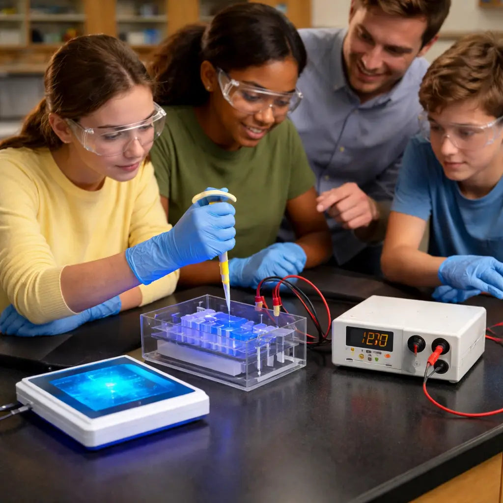
Picture this: you’re setting up a high‑school biology lab, the bell rings, and you’ve got a class of eager teens ready to see DNA fragments separate on a gel. The excitement is real, but the budget spreadsheet is screaming ‘no.’
Does a "budget friendly gel electrophoresis system for classrooms" sound like a myth? Not at all. The trick is to focus on what actually matters for teaching—reliability, ease of use, and cost‑effectiveness—without getting lost in fancy specs you’ll never need.
First, think about the power supply. A compact, low‑voltage unit that runs on a standard outlet can cost a fraction of the industrial‑grade models. Look for built‑in safety features like automatic shut‑off; they keep curious hands safe and let you spend the saved dollars on consumables.
Next up, the gel tank. A lightweight acrylic tank with a simple gel casting system is perfect for a classroom. You don’t need a massive cooling system—just a decent buffer reservoir and a clear lid so students can watch the bands form in real time.
And what about the transilluminator? LED‑based blue‑light units are now under $150 and eliminate the UV hazards that make teachers nervous. Pair that with a low‑cost, high‑contrast agarose—something you can reuse a few times before discarding—to stretch every penny.
In our experience at Shop Genomics, we see educators gravitate toward bundled kits that include a power supply, tank, and LED transilluminator. The bundles shave off about 15% compared to buying each piece separately, and the free‑shipping threshold on larger orders helps keep the total landed cost down.
So, how do you turn this into a doable plan? Start by listing the essential components: power supply, tank, transilluminator, and a basic set of pipettes. Then compare prices across vendors, but give priority to bundles that bundle the three core pieces. Remember, the goal isn’t to get the flashiest setup—it’s to give students a hands‑on moment they’ll remember when they later see a DNA fingerprint in a news story.
Finally, don’t forget the little things that make a big difference: a sturdy bench‑top stand to keep the tank steady, a set of reusable loading tips, and a simple ladder marker that’s easy to read. When you line these up, you’ve built a classroom‑ready gel electrophoresis system that fits snugly into a tight budget while still delivering clear, teachable results.
TL;DR
You can set up a budget‑friendly gel electrophoresis system for classrooms by picking a compact power supply, a lightweight acrylic tank, and an LED blue‑light transilluminator, then bundling them for up to 15 % savings and reusing agarose gels a few times.
This approach gives high‑school students clear, safe visual results without breaking the school’s budget, and you’ll have a reusable, safe setup that works semester after semester.
Choose the Right Low‑Cost Electrophoresis Kit
When you’re pulling together a budget friendly gel electrophoresis system for classrooms, the biggest headache is figuring out which pieces actually matter. You’ve already settled on a compact power supply, a lightweight acrylic tank, and a blue‑light transilluminator. Now it’s time to match those parts so they work together without blowing the budget.
First thing’s first: know what you need in the lab. Ask yourself, “Do my students only run 100–1,000 bp PCR fragments, or will they also look at larger plasmid digests?” The answer tells you how much agarose you’ll need and whether a 0.8 % gel will do or you’ll need a 2 % mix for sharper bands. It also guides the voltage range you’ll set on the power supply.
Power supply – keep it simple and safe
Low‑cost units that plug straight into a standard outlet are perfect for high‑school benches. Look for a model with an automatic shut‑off and a clear voltage read‑out. That way you avoid the dreaded “over‑run” that leaves everyone with smeared bands and a nervous teacher.
Pro tip: set the voltage according to the rule of 5–10 V per centimetre of gel length. If you’re running a 10 cm gel, 60 V gives you a nice, steady migration without heating the gel too fast.
Tank – size does matter, but not in the way you think
Acrylic tanks with a removable lid are cheap, lightweight, and let students peek at the bands as they separate. You don’t need a cooling system; just make sure the buffer reservoir is large enough to keep the electrodes submerged throughout the run.
If you’re buying a kit, compare the tank’s internal dimensions to the size of the comb you’ll use. A snug fit means less buffer waste and a more stable gel.
Transilluminator – go blue, skip UV
LED blue‑light units are the safest choice for classrooms. They’re usually under $150 and give you the high‑contrast images you need for grading lab reports. The only thing to watch is the filter – a green filter works best with SYBR Safe or GelRed stains.
When you’re setting up the imaging station, think about the workflow. A bench‑top stand that tilts a few degrees makes it easier for students to snap a quick photo without bending over the bench.
Bundle for savings
Many suppliers, including Shop Genomics, offer bundled kits that pair the power supply, tank, and transilluminator. Bundles shave off roughly 15 % compared to buying each piece separately, and you often qualify for free shipping on larger orders.
But don’t stop at the big three. Adding a set of reusable loading tips, a simple ladder marker, and a basic pipette set rounds out the kit without adding much cost.
Need a way to keep the prep time tight? Try using a Pomodoro timer to break the gel‑casting and loading steps into focused 25‑minute bursts. It keeps students on track and reduces the chance of forgetting to add the buffer. You can read a quick guide on distraction‑free coding sprints that works just as well for lab work here.
Don’t forget the paperwork. Printing ladders, sample labels, and safety signs can be done on a low‑cost online printer service. A fast, reliable print shop that ships quickly can save you a trip to the campus copy centre. Check out Jiffy Print Online for affordable, on‑demand prints.
Once you’ve gathered all the pieces, do a quick “dry run” with water instead of buffer. Make sure the electrodes line up, the tank sits level, and the transilluminator lights up without flicker. If anything feels off, swap the component before you pour the agarose – it’s way easier to fix a mistake at this stage than after the gel’s set.
Now that you’ve seen the kit in action, it’s time to put it together. Follow the checklist below before you head to the next class:
- Confirm voltage range matches gel length.
- Check tank dimensions against comb size.
- Verify transilluminator filter matches stain.
- Pack reusable tips and ladder markers.
- Print labels and safety signs ahead of time.
With those steps checked off, you’ll have a reliable, low‑cost electrophoresis setup that lets students focus on the science, not the price tag.
Set Up a Simple Power Supply
Okay, you’ve got the tank and the blue‑light viewer ready—now it’s time to bring the power back. A modest, budget friendly gel electrophoresis system for classrooms can run off a tiny 12‑V, 300‑mA supply that plugs into any standard outlet. No fancy racks or coolant loops needed.
Pick the right little box
First, look for a supply that’s labeled “compact” or “bench‑top” and has an automatic shut‑off. Those safety switches are a lifesaver when curious hands wander over the bench. If the unit shows a voltage range of 0‑12 V and a current limit around 300‑500 mA, you’re in the sweet spot for a 10‑cm agarose gel.
In our experience at Shop Genomics, the 12‑V, 300‑mA model we stock consistently hits that budget‑friendly sweet spot without compromising safety.
Unbox and inspect
When you pull the power supply out of the box, give it a quick visual check. Make sure the plug isn’t frayed, the knobs turn smoothly, and the display (if there is one) reads clearly. A cracked case or missing knob can turn a simple run into a pricey repair.
Tip: keep the unit on a non‑conductive stand—something as simple as a small plastic rack will keep the bench tidy and protect the supply from accidental spills.
Wire it up the easy way
Most low‑cost supplies come with two banana‑plug leads: red for positive (anode) and black for negative (cathode). Snap the red lead into the “+” terminal on the supply, the black into “–”. Then plug the other ends into the electrophoresis tank’s electrode clips. If your tank has built‑in clips, just press the plugs onto them; you’ll hear a soft click.
Don’t force anything. If a plug won’t seat, double‑check you’re using the right polarity—mixing them up can fry the gel and the supply.
Set the voltage, then watch the dye front
Turn the dial to 50‑60 V for a 10‑cm gel. That’s enough to move the loading dye about three‑quarters of the way down in roughly 30‑45 minutes. You’ll know the run is on track when the orange bromophenol blue line crawls past the halfway mark.
Here’s a quick sanity check: if the dye rushes to the bottom in under 20 minutes, dial the voltage back a notch. If it crawls slower than 1 cm per minute, bump it up a little.
Safety first, always
Never touch the electrodes while the power is on—keep your hands dry and wear rubber‑soled shoes. If a student asks, explain that the current is invisible but can still give a nasty shock.
When the dye front reaches the ¾ point, hit the off switch. Many supplies have a “stop” button that cuts power instantly; if yours only has a dial, turn it down to zero.
Test run checklist
- Supply plugged into a grounded outlet.
- Red lead in the anode, black in the cathode.
- Voltage set between 50‑60 V for a standard 10‑cm gel.
- Timer set for 30‑45 min, watch the dye front.
- Automatic shut‑off engaged.
If everything looks good, you’re ready for the real samples. If the gel looks fuzzy or the power flickers, double‑check the connections and make sure the buffer fully covers the electrodes.
Quick troubleshooting
Loose plug? Re‑seat it and listen for a solid click. Power supply humming but voltage won’t rise? The unit may be in “current limit” mode—lower the load by using a fresh buffer or a slightly lower voltage.
Still stuck? A short video from Ward’s World walks through the basics of classroom gel electrophoresis tools and shows how a simple power supply fits into the whole workflow (see the guide for tips). It’s a handy visual reminder that the setup isn’t rocket science.
Once you’ve mastered the supply, the rest of the experiment feels almost automatic. You’ll spend less time fiddling with wires and more time watching those bright bands light up the students’ faces.
So, what should you do next? Grab your power supply, follow the checklist, and run a test gel with just the loading dye. When that first run looks clean, you’ll know your budget‑friendly system is good to go for the whole semester.
Assemble the Gel Casting Tray
Alright, you’ve got your power supply and tank ready – now it’s time to build the heart of the system: the gel casting tray. If you’ve ever tried to force‑fit a commercial tray that cost $80‑$120 into a tight budget, you know the frustration. The good news? You can make a perfectly good tray for a fraction of the price with a few sheets of acrylic and a little CAD know‑how.
Gather the right pieces
Start with a 6 mm thick clear acrylic sheet for the base – it’s sturdy enough to hold the gel and cheap enough to cut yourself. A 1.5 mm acrylic strip works great for the side walls; you’ll need four pieces that match the length of your tank (usually 12 cm for a 10‑cm gel). If you have a laser cutter in the department’s maker space, great. If not, a handsaw and a file will do; just take your time to keep the edges smooth.
Don’t forget the comb. You can buy a cheap plastic comb for $5, but custom‑designed combs cut from 1.5 mm acrylic give you exactly the well spacing you need – whether you’re loading 8, 12, or even 96 wells. Dr Chris Pook walks through a simple OpenSCAD design that you can tweak in under ten minutes (custom tray design guide).
Cut and assemble the base
Measure twice, cut once. Mark the dimensions on the 6 mm sheet: length, width, and a shallow groove (about 2 mm deep) along the back edge where the side walls will sit. Use a fine‑toothed saw or a laser cutter to make the cuts, then sand the edges with 120‑grit sandpaper so they don’t chip the acrylic.
Slide the four side walls into the groove. If the fit is a hair loose, add a dab of clear silicone sealant at each corner – it’s cheap, non‑conductive, and dries clear. Let it cure for an hour before moving on.
Make the comb
Take the 1.5 mm strip and lay it flat on a piece of cardstock. Sketch the comb teeth – 6 mm wide and 5 mm tall is a comfortable size for most 1 mm‑deep gels. Cut the teeth with a razor blade or a small rotary cutter. The teeth should be evenly spaced; a ruler or a simple 3D‑printed spacer can help keep things consistent.
Test the comb by inserting it into a test gel (you can use a tiny 5 ml petri dish). If the teeth bend, thin the walls a touch. If they don’t reach the bottom, shave a millimetre off the base of each tooth.
Test‑fit the tray in the tank
Place the assembled tray into your acrylic tank. The bottom should sit flat, and the walls should be flush with the tank’s inner edges. Fill the tank with running buffer – just enough to cover the electrodes but not so much that it overflows when you pour the agarose.
Pour a thin “test gel” (0.5 % agarose) and let it set. Once solid, gently lift the tray; the gel should peel away cleanly, and the comb should snap out without tearing. If the gel sticks, a light coating of petroleum jelly on the tray walls does the trick.
Pro tips for a budget‑friendly system
- Re‑use the same acrylic tray for up to ten runs. Just wash with distilled water and a mild detergent between runs.
- Keep a spare set of comb teeth cut from the same strip – they’re cheap to replace and prevent a broken comb from ruining a whole class.
- When you’re ready to scale up, design a modular tray that slides into a larger 15‑cm tank; the same side walls work, you just add a longer base.
In our experience at Shop Genomics, teachers who build their own trays report a 30 % reduction in consumable cost compared with buying pre‑made kits. Plus, the hands‑on building step becomes a mini‑engineering lesson for the students.
Need a quick price‑check on the tank you’ll be pairing this tray with? See our guide on understanding gel electrophoresis tank pricing for budgeting tips.
Finally, if you want to keep track of each run – which DNA samples were loaded, voltage used, and buffer changes – a simple lab‑management tool can save you hours of paperwork. For example, the compliance‑focused platform ClientBase (https://clientbase.pro) offers a free tier that’s perfect for high‑school labs.
Compare Popular Budget Gel Systems
When you’re trying to stretch a tight classroom budget, the gel electrophoresis system you pick can make or break the whole semester. Let’s look at three ways teachers have gotten reliable, hands‑on DNA runs without blowing the budget.
1. DIY acrylic tray + low‑cost power supply
Think of this as the "build‑your‑own" option. You buy a 12‑V, 300‑mA bench‑top supply (often under $80), a lightweight acrylic tank, and cut your own tray from a sheet of clear acrylic. The upfront cost can be under $120 total, and you can reuse the tray for ten runs or more.
Pros: total control over dimensions, easy to repurpose for other experiments, and the hands‑on construction becomes a mini‑engineering lesson. Cons: you need a bit of shop‑class skill and a few extra minutes for assembly.
What we’ve seen work best is pairing the DIY setup with a cheap reusable comb and a simple blue‑light viewer you can snag for $30‑$40. The whole rig stays under $200, which leaves room for consumables.
2. miniPCR Bandit™ + Viewit™ kit
The miniPCR Bandit™ + Viewit™ kit bundles a reusable electrophoresis chamber, a compact power module, and an illumination unit that lets students watch DNA separate in real time. Priced at about $150, it’s a true all‑in‑one solution.
Pros: plug‑and‑play out of the box, safe low‑voltage design, and the Viewit™ illumination eliminates the need for a separate transilluminator. Cons: you’re locked into the kit’s dimensions (good for 10‑cm gels) and you can’t customize the tray beyond what’s supplied.
Teachers love that the kit is fully reusable—no disposable trays to replace each semester—and the price still fits comfortably under most department budgets.
3. MiniOne all‑in‑one bundle
MiniOne packages a power supply, acrylic tank, blue‑light viewer, and even a micropipette into a single rugged case. The price hovers around $199, so it’s a shade pricier than the Bandit combo but still well below commercial‑grade systems.
Pros: everything arrives pre‑assembled, so you can set up in under ten minutes and get straight to the experiment. Cons: the bundled micropipette may not be ideal for labs that already own their own tips, and the case adds a little bulk.
If you have a busy schedule and need a system that can be grabbed off the shelf, MiniOne is the safest bet.
Quick side‑by‑side comparison
| System | Approx. Cost | Key Features | Best For |
|---|---|---|---|
| DIY acrylic tray + power supply | $120‑$150 | Customizable tray, reusable components, low‑voltage safety | Classes that enjoy a hands‑on build project |
| miniPCR Bandit™ + Viewit™ | $150 | All‑in‑one kit, real‑time illumination, reusable gel chamber | Teachers who want a plug‑and‑play solution |
| MiniOne bundle | $199 | Pre‑assembled, includes pipette, compact blue‑light viewer | Fast‑setup labs with limited assembly time |
So, which one feels right for your classroom? If you have a maker‑space and want students to see how the tray is built, the DIY route gives you flexibility and a learning moment. If you’d rather spend class time looking at bands rather than tightening screws, the Bandit+Viewit kit slides in perfectly. And if you need a system that’s ready to roll the minute you open the box, MiniOne saves you the extra prep.
One final tip: whatever system you choose, write down the exact model, voltage setting, and run time in a simple spreadsheet. That habit turns a one‑off experiment into a repeatable protocol you can hand to the next teacher, and it keeps your budget‑friendly gel electrophoresis system for classrooms humming year after year.
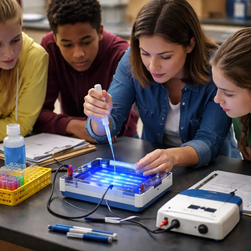
Run Your First Sample and Interpret Results
Alright, you’ve got your power supply, tank, and gel tray all set up. The moment of truth is loading that first sample and watching the DNA dance across the gel. It feels a bit like a science‑fair magic trick, doesn’t it?
Step 1 – Prep your samples
First, mix your PCR product (or any DNA fragment) with a loading dye. The dye does two things: it adds weight so the sample sinks into the well, and it lets you track the migration front. A common recipe is 1 µl dye to 9 µl sample – keep it simple and you’ll avoid over‑loading.
Tip: If you’re teaching a class of 20‑plus students, label each tube with a colour‑coded sticker. It turns a bland lane into a story the kids can follow.
Step 2 – Load the gel
Grab a pipette, set the volume to match your well depth (usually 5‑10 µl), and gently push the tip to the bottom of the well. Release the sample slowly – a steady drip looks cooler than a splash, and it prevents bubbles.
And what about the ladder? Always load a size marker in the first lane. It’s the ruler that tells you, “Hey, that bright band is about 500 bp.”
Step 3 – Start the run
Turn the voltage knob to 50‑60 V for a 10‑cm gel. You’ll see the orange bromophenol blue dye front crawl about three‑quarters of the way down in 30‑45 minutes. When it hits that sweet spot, hit the off switch.
In our experience, a run that finishes too fast (under 20 minutes) usually means the voltage was too high – the bands will look smeared. Too slow, and you waste class time.
Step 4 – Visualize the bands
After the power is off, remove the gel and place it on your blue‑light transilluminator. If you’re using the miniPCR budget‑friendly classroom kit, the Viewit™ viewer lets the whole class see the bands in real time – no UV, no fuss.
Adjust the camera exposure until the faintest band is just visible. Too bright and the ladder loses detail; too dim and you’ll miss low‑abundance fragments.
Step 5 – Interpret the results
Now the fun part: reading the gel. Compare each sample lane to the ladder. If a band lines up with the 500 bp marker, you’ve got a 500‑base‑pair fragment. Multiple bands? That could mean a mixed PCR product or non‑specific amplification – perfect teachable moments.
Ask your students: “What does it mean if a band is fainter than the ladder?” They’ll discover that lower intensity often signals less template or incomplete staining.
When you spot an unexpected band, note it in a simple spreadsheet: lane number, expected size, observed size, intensity (bright, medium, faint), and any odd observations. This habit turns a one‑off run into a repeatable protocol you can hand to the next teacher.
Quick checklist before you call it a day
- Samples mixed with loading dye (1:9 ratio).
- Ladder loaded in first lane.
- Voltage set to 50‑60 V for a 10‑cm gel.
- Dye front reached ~¾ of the gel length.
- Transilluminator turned on, exposure adjusted.
- Results logged in a spreadsheet.
And if something went sideways – a fuzzy band, a missing lane, or a broken tip – don’t panic. Grab a fresh tip, double‑check the buffer level, and run a quick test gel with just the loading dye. Most hiccups are easy to fix, and the class learns problem‑solving on the spot.
So, what should you do next? Run that first sample, snap a photo of the gel, and write down the key observations. The next time you pull out the same budget‑friendly gel electrophoresis system for classrooms, you’ll be ready to hit the ground running and let the students see DNA come alive.
Additional Resources & Printable Guides
When you’ve nailed the basic run, the next step is to give yourself and your students a set of ready‑to‑print guides that take the guesswork out of every gel.
We’ve pulled together a handful of PDFs that cover everything from buffer preparation to ladder selection, all formatted for a quick printout that fits on a standard 8 × 11 sheet.
If you prefer a step‑by‑step video‑free cheat sheet, the University of Wisconsin’s molecular ecology lab protocol is concise, downloadable and perfect for labs on a tight schedule.
miniPCR teaching toolkit also offers free worksheets, sample data sets and a troubleshooting flowchart – a handy companion when you’re introducing electrophoresis to a new cohort.
Tip: print the buffer‑mix table on cardstock so it stays flat on the bench, and laminate the ladder‑size chart – a quick glance saves a minute of scrolling on a phone.
And if you like to keep a digital record, create a shared folder on your school’s drive and drop each PDF in there; the whole department can pull the same resources for every semester.
Finally, bookmark these guides in your lab notebook – when a new teacher steps in, they’ll have a ready‑made roadmap to keep the gel runs smooth and the students engaged.
Conclusion
We've walked through everything you need to pull off a budget friendly gel electrophoresis system for classrooms, from picking the right power supply to printing cheat‑sheet PDFs.
So, what’s the biggest win? You can give students a real DNA‑separation moment without draining the department budget, and you still have a setup that lasts semester after semester.
Remember to keep the core components – low‑voltage supply, acrylic tank, blue‑light viewer, reusable comb – on a shared drive or a laminated card. That tiny habit saves minutes every class.
In our experience at Shop Genomics, teachers who bundle the essentials and label each part see fewer hiccups and more time for data discussion.
Before you close the lab for the day, run a quick checklist: voltage set, buffer topped up, ladder printed, and a spare tip rack within reach.
Does this feel doable? Absolutely. The pieces are inexpensive, the instructions are printable, and the science is eye‑opening.
Next step: pick one of the ready‑to‑print guides we highlighted, print it on cardstock, and slot it into your lab notebook. Tomorrow’s class will thank you.
And if you ever need a fresh quote or a new kit, platforms like Shop Genomics make ordering painless, with free shipping on larger orders and easy returns.
FAQ
How can I keep a budget friendly gel electrophoresis system for classrooms running smoothly over multiple semesters?
First, label every core part – power supply, tank, LED viewer, comb – and stick the list on a laminated card. That tiny habit saves minutes when you set up each class. Second, store the acrylic tray flat and wipe it with distilled water after each run; a quick dry prevents mold and keeps the gel surface clean. Finally, run a short “test gel” with just loading dye every few weeks; it catches loose connections before a student experiment starts.
What voltage settings are safe for a low‑cost power supply in a high‑school lab?
For a standard 10‑cm gel, aim for 50‑60 V. That range moves the bromophenol blue front in about 30‑45 minutes without overheating the agarose. If you notice the dye rushing past the halfway mark in under 20 minutes, dial the voltage down a notch. Keep the supply on automatic shut‑off mode so it cuts power if the current spikes – a simple safety net that costs nothing.
How do I choose the right agarose concentration without overspending on consumables?
Match the gel % to the fragment size you expect. Around 1 % works well for 300‑800 bp PCR products, while 0.8 % gives clearer separation for larger pieces. Buy agarose in bulk packs; the per‑gram price drops dramatically and you can store leftovers in a dry cabinet for months. When you’re unsure, start with a 1 % gel – it’s the most forgiving middle ground.
Can I reuse the gel tray and comb, and how should I maintain them?
Absolutely. The acrylic tray can survive ten or more runs if you rinse it with distilled water and a mild detergent after each experiment. For the comb, a quick soak in 70 % ethanol removes residual DNA and prevents cross‑contamination. Inspect the teeth for chips before each use; a broken tooth can ruin an entire lane, so keep a spare set of teeth cut from the same acrylic strip.
What are the best practices for storing and preparing running buffer on a tight budget?
Make a large batch of 1× TBE or TAE and store it in a sealed container at room temperature – the buffer stays good for six months if you keep the lid tight. Before each run, top up the tank so the electrodes stay submerged but don’t overfill; excess liquid can spill onto the bench and damage equipment. If the buffer looks cloudy, filter it through a 0.22 µm syringe filter rather than buying a new bottle.
Where can I find printable cheat‑sheet guides that fit a budget friendly gel electrophoresis system for classrooms?
Shop Genomics offers a collection of free PDFs that cover everything from buffer recipes to voltage checklists. You can download them, print on cardstock, and laminate for repeat use. Having the cheat‑sheet on the bench means you and your students spend less time hunting for numbers and more time watching the bands appear. A quick glance at the guide also reminds you of safety steps before you power up.


